Gene Fusion Probe
Due to chromosomal rearrangement, a fusion gene formed by joining two parental genes is a hallmark of many types of cancer. Structural gene fusion rearrangements that cause abnormal signals are frequently detected in many cancer types. When two normally separated genes are joined, they will fuse and promote the development of cancer in several different ways. When a translocation occurs, the gene can be fused with a promoter to increase gene expression. Due to chromosomal rearrangement, the fusion formed by joining two parental genes can also produce chimeric genes. The new fusion gene produces a new hybrid protein with malignant consequences. In addition, when the fusion causes the tumor suppressor gene to be truncated, it can lead to tumorigenesis. Gene fusion probes can be used to detect common translocations involving gene regions that can cause leukemia and/or cancer when rearranged and connected.
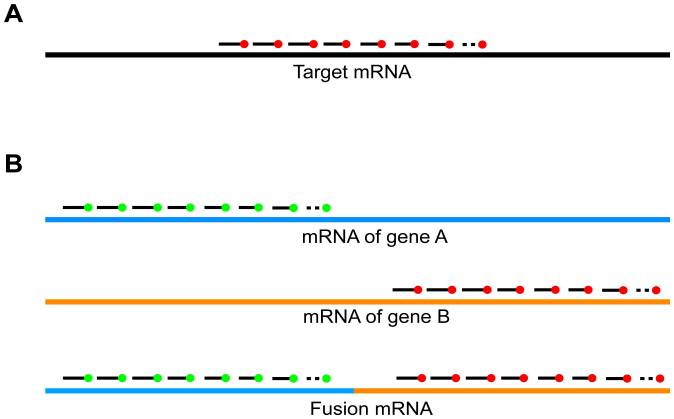 Fig 1. Principle of Fusion FISH imaging. (Markey F B, et al. 2014)
Dual-color Gene Fusion Probe Customization Services
Gene fusion has important prognostic and predictive value and may be used as a molecular marker for molecular pathology tests for patient management. Many molecular biology methods are used to identify specific fusion genes, among which the FISH method can visualize the presence of fusion genes on chromosomes. Double fusion probes are used to detect specific translocations related to malignant biological events such as cancer. The analysis of fusion transcripts in cancer and other special pathological samples can be performed by dual-color probes designed separately for fusion genes. Each color is designed to cross with its own gene. Normal cells will display colors as separate signals, while cells with fusion will display signals separated by less than one signal width. Our service can design the sequence of the target gene submitted by the customer, prepare two sets of probe sets of different colors, and label them with different fluorescent dyes to form a convenient tool for detecting fusion genes. The customization of our two-color gene fusion probes is done by professional scientists, and the sequence can be retrieved from public databases or the sequencing data submitted by customers. At the same time, we provide a variety of fluorescent dyes and markers to provide customers with more fluorescent detection possibilities.
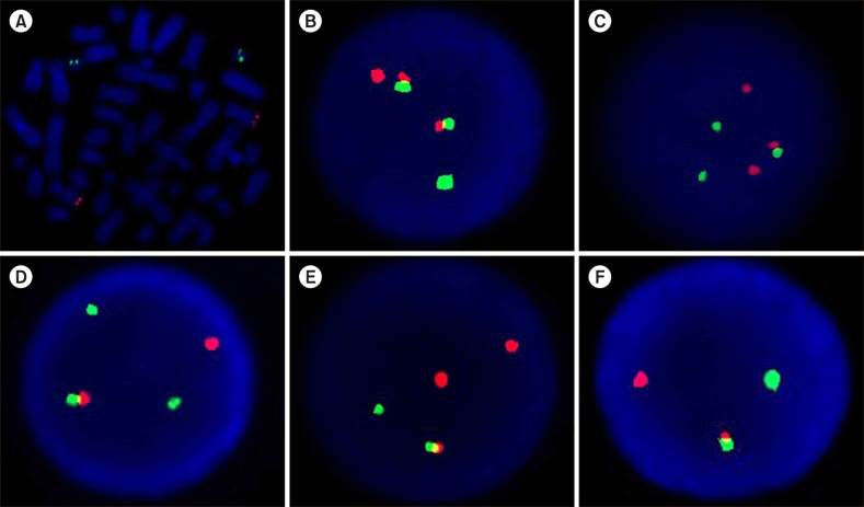 Fig 2. Representative FISH signal patterns using LSI BCR/ABL dual color dual fusion probe. (Švabek Ž T, et al. 2018)
Creative Bioarray provides a customized service of two-color FISH probes for detecting chromosome translocation and gene fusion. This service can be used for chromosome visualization analysis of genetically related diseases such as cancer. You will benefit from our internally optimized probe design scheme, which uses two colors to label different target genes. We aim to provide you with a cost-effective custom solution for fusion FISH probes. If you are interested in our probe customization service, please contact us for customization. We look forward to cooperating with you in the near future.
References
- Markey F B, Ruezinsky W, Tyagi S, et al. Fusion FISH imaging: single-molecule detection of gene fusion transcripts in situ[J]. PloS one, 2014, 9(3): e93488.
- Solomon J P, Hechtman J F. Detection of NTRK fusions: merits and limitations of current diagnostic platforms[J]. Cancer research, 2019, 79(13): 3163-3168.
- Švabek Ž T, Josipović M, Horvat I, et al. The incidence of atypical patterns of BCR-ABL1 rearrangement and molecular-cytogenetic response to tyrosine kinase inhibitor therapy in newly diagnosed cases with chronic myeloid leukemia (CML)[J]. Blood research, 2018, 53(2): 152-159.
All products and services on this website are only suitable for non-medical purposes.


