Prokaryotic Cells
Creative Bioarray provides customized in situ visualization solutions for analyzing gene expression and localization in prokaryotic cells. Our FISH technical services can help customers achieve quantitative or qualitative target mRNA in prokaryotes in biological samples and non-biological environmental samples. At the same time, our molecular platform also provides related protein level analysis services, such as IHC analysis of corresponding targets. Diversified choices, high-quality service teams, and complete equipment platforms give us the confidence to provide customers with comprehensive analysis and testing services.
Prokaryotic cells and FISH technology
In recent years, the demand for visual analysis of prokaryotic cells in various fields such as environment, food, and medicine has gradually increased. FISH technology facilitates the analysis of microorganisms in complex samples by using fluorescently labeled nucleic acid probes. FISH with rRNA-targeted oligonucleotide probes has become one of the core technologies in microbial ecology today, which enables in situ identification and quantification of microorganisms and prokaryotic cells in environmental samples. Classic FISH protocols feature the use of fluorescent single-labeled probes. To cope with the various difficulties of prokaryotic analysis, more sensitive TSA-FISH or CARD-FISH techniques involving enzymatically catalyzed signal amplification steps have been developed on the basis of classical protocols. Classical FISH has the advantage of being relatively inexpensive and easy to operate on morphologically diverse samples. TSA-FISH and CARD-FISH offer significantly higher sensitivity, allowing the detection of slow-growing or metabolically inactive cells below the detection limit of classical FISH. The accuracy of FISH analysis has been improved through multiple probes and optimized cell fixation and permeabilization methods. Our experimental platform provides customers with customized FISH solutions for the visualization and analysis of prokaryotic cells.
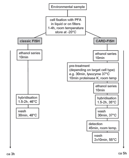 Flow chart showing steps for classic and CARD-FISH. (Zwirglmaier K, et al. 2010)
Prokaryotic Cell Visualization Analysis Solution
The analysis method of FISH was mostly used for the analysis of eukaryotes in the early days, and later evolved a scheme suitable for prokaryotes. We offer customization of prokaryotic FISH analysis solutions based on TSA-FISH or CARD-FISH technology. At present, FISH analysis of prokaryotes can be widely used in the analysis of pathogens in biological samples, or the study of microorganisms in environmental samples from non-living organisms. We provide custom solutions for customers with prokaryotic gene expression and localization analysis needs, which are designed according to your sample type and analysis purpose. And choose suitable probe solutions for customers, you can choose existing commercial probes or customize them.
Our technical services can fulfill the following application analysis needs:
Using ribosomal RNA as a target provides direct quantification of living cells defined by the presence of ribosomes;
Qualitative or existential analysis of prokaryotes in abiotic environmental samples;
To corroborate and quantify by other quantitative methods such as QPCR;
Features of FISH Visual Analysis of Prokaryotic Cells
- Variants of FISH technology can analyze prokaryotic cells in biological samples, as well as in situ identification and quantification of microbial cells in ecosystems;
- rRNA is an ideal target for observing distant cells;
- 16S rRNA has a large and growing database of available sequences (it has become the most common target for FISH probes);
- rRNA serves as a phylogenetic marker of prokaryotic cells and is present in metabolically active cells in high copy numbers;
- Increased sensitivity through multiplex TSA-FISH methods;
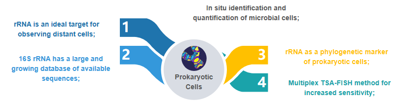 Fig 2. Availability of FISH assays for prokaryotic cells.
If you are interested in our service, please contact us for cooperation. We look forward to cooperating with you in the near future.
References
- Zwirglmaier K. Detection of prokaryotic cells with fluorescence in situ hybridization[M]//Fluorescence in situ Hybridization (FISH). Humana Press, Totowa, NJ, 2010: 349-362.
- Wilhartitz I, Mach R L, Teira E, et al. Prokaryotic community analysis with CARD‐FISH in comparison with FISH in ultra‐oligotrophic ground‐and drinking water[J]. Journal of applied microbiology, 2007, 103(4): 871-881.
- Kawakami S, Hasegawa T, Imachi H, et al. Detection of single-copy functional genes in prokaryotic cells by two-pass TSA-FISH with polynucleotide probes[J]. Journal of microbiological methods, 2012, 88(2): 218-223.
All products and services on this website are only suitable for non-medical purposes.


