HPV Integration
HPV Integration and Oncogenesis
Human papillomavirus (HPV) is a DNA virus that infects the skin and mucous membranes and contains more than 100 types. Studies have classified HPV-related carcinogenesis. About one-fifth of HPV has a high risk of multiple cancers (including HPV 16, 18, 31, 33, etc.). HPV has a small double-stranded circular DNA genome of approximately 7-8 kb in size. It is usually present in the nucleus of infected cells in the form of circular extrachromosomal elements, but may accidentally integrate into the host chromosome. HPV integration can occur as a single integrated virus, called type I integration, or multiple tandem integrated viruses, called type II integration. In the HPV integration site with amplified HPV DNA, the host DNA can be co-amplified to produce a unique HPV host concatemer, called the type III integration site. Fluorescence in situ hybridization (FISH) is a common technique used to visualize and locate HPV DNA in HPV-infected cells and tissue sections cultured in vitro. Limited by resolution, interphase FISH is not available in HPV analysis, while interphase FISH is used to distinguish individual HPV integration sites. Fiber-FISH (Fiber-FISH) can improve the resolution of analyzing HPV integration sites, and can be used to detect and distinguish type I, II, and III integration, and determine the co-amplification mode of HPV hosts.
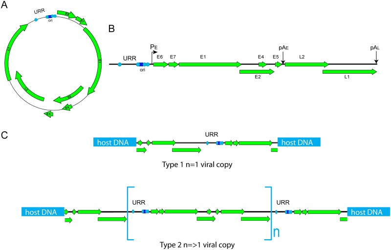 Fig 1. Types of HPV integration. (McBride A A, et al. 2017)
FISH Service for HPV
We provide FISH test services that can be used for HPV analysis in research projects related to cancer occurrence-related HPV integration events. This service can analyze fixed cell and tissue samples. We use interphase chromosome preparation and high-resolution imaging. The basic processes of these services include probe customization, sample preparation, chromosome preparation (chromosome fiber preparation), FISH hybridization, imaging, and data acquisition. Our optimized Fiber-FISH experimental protocol can be used to analyze the type of HPV integration and visualize the integration site. In addition, our service can distinguish integrated and non-integrated targets using chromosome-specific probes. The resolution of mid-term FISH can distinguish individual HPV integration sites.
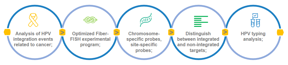 Fig 2. FISH analysis of HPV integration events.
Some potential HPV integration events that can be used as detection targets
- Disruption of E2 gene
- Disruption of E2 binding sites by methylation
- Disruption of E1 gene
- Formation of viral—host fusion transcript
- Tandem repeats of HPV genome/regulatory elements
- Altered regulation of cancer-associated genes in the vicinity of the integration locus
Creative Bioarray provides FISH analysis services for HPV in cancer research. This service can use mid-term FISH analysis and Fiber-FISH to analyze the type of HPV integration and visualize the integration site. This service can be carried out for cell and tissue samples, and you will benefit from our internal optimized technology portfolio. If you are interested in our FISH solution for HPV integrated analysis, please contact us for cooperation. You will experience a one-stop service and look forward to cooperating with you in the near future.
References
- McBride A A, Warburton A. The role of integration in oncogenic progression of HPV-associated cancers[J]. PLoS pathogens, 2017, 13(4): e1006211.
- Redmond C J, Fu H, Aladjem M I, et al. Human Papillomavirus Integration: Analysis by Molecular Combing and Fiber‐FISH[J]. Current protocols in microbiology, 2018, 51(1): e61.
All products and services on this website are only suitable for non-medical purposes.


