Three-dimensional Analysis and Visualization of Chromosome Conformation
Different methods for visualizing genome topology and spatial relationships between genes have received extensive attention as tools for decoding the relationship between chromatin structure and function. 3D-FISH on cultured cells has become a routine technique that uses fluorescently labeled nucleic acid probes to detect the part of the genome in the nucleus of metaphase or interphase cells and is now widely used in nuclear biology. This technology can visualize chromosomal regions, chromosomal subregions, individual genes, and RNA transcripts, thereby preserving their spatial position in the nucleus. In some specific cases, it is necessary to combine 3D-FISH and immunostaining to map DNA/RNA and protein targets in the same cell. FISH can be combined with the detection of nuclear proteins, but the difficulty lies in the possible interference between the FISH procedure and immunostaining, such as differences in antigen stability and immunostaining molecular tools (antibodies). Our combined service solutions can be customized according to the needs and goals analyzed by customers.
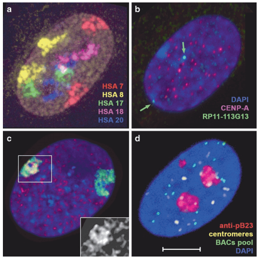 Fig 1. Examples of 3D-FISH and 3D-FISH combined with immunostaining. (Solovei I, et al. 2010)
FISH Services Related to Nucleic Acid Analysis
Our 3D-FISH uses DNA probes to perform fluorescence in situ hybridization on 3-dimensionally preserved cell nuclei, and then visualize the location of gene loci, chromosomal subregions or the entire region in a single cell through a 3D confocal microscope. Customers can use this direct method to understand the overall structure of the cell nucleus and the behavior of specific genomic sites and regions in nuclear space. On this basis, we provide immunostaining methods to detect nuclear proteins, including modified histones, histone variants and modifications, transcription mechanisms and factors, and nuclear compartments. According to the FISH guidelines, we provide customized solutions based on differences in protein target stability to help customers solve the dilemma of simultaneous analysis.
This service is carried out for primary cell samples separated and digested from cultured cells. Our service includes the steps of sample pre-processing and can provide technical support from the beginning of cell harvesting. According to the stability of the antigen to be tested (determining the staining order) and the different staining methods (whether it is a fluorescently labeled antibody), we provide different staining schemes. The standardized tests of these programs have been verified on our experimental platform, and our experienced experimenters can provide full experimental records. In addition, our microscope platform has a variety of models of microscopes, providing multi-channel and high-resolution image acquisition. Finally, our experimental team can perform further analysis on the collected images.
Methodology and Customized Solutions
The main challenge in combining immunofluorescence or anti-biotin-conjugated antibody-mediated immunostaining methods with 3D DNA FISH is to retain the 3D structural analysis of the epitope and nucleus detected by the antibody. We provide detection strategies based on FISH guidelines to stain antigens. For stable antigens, antibody binding can be performed after fixation, and stable antigens can be performed after FISH hybridization. Our technical services will provide customized solutions for the antigens you are interested in.
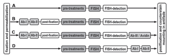 Fig 2. The basic workflow of the four typical variants of the 3D-FISH and 3D-immuno-FISH protocols. (Solovei I, et al. 2010)
Creative Bioarray provides immunostaining and 3D-FISH technical services to decode the relationship between chromatin structure and function, helping our customers analyze DNA/RNA and protein targets in the same cell at the same time. According to the differences in the stability of protein targets and the differences in molecular tools, we develop the most suitable solutions for customers. If you are interested in our Immunostaining and 3D-DNA FISH services, please contact us for cooperation. We look forward to cooperating with you in the near future.
References
- Solovei I, Cremer M. 3D-FISH on cultured cells combined with immunostaining[M]//Fluorescence in situ Hybridization (FISH). Humana Press, Totowa, NJ, 2010: 117-126.
- Chaumeil J, Micsinai M, Skok J A. Combined immunofluorescence and DNA FISH on 3D-preserved interphase nuclei to study changes in 3D nuclear organization[J]. Journal of visualized experiments: JoVE, 2013 (72).


