Heterochromatin-Directed Multicolor FISH
Heterochromatin Visualization and Multicolor FISH
Methods for the detailed characterization of chromosomal rearrangements detected in conventional band cytogenetics have been developed using FISH and/or array comparative genomic hybridization (aCGH). Compared to the higher resolutions available in aCGH, FISH has the advantage that it allows the analysis of balanced rearrangements and chromosomal aberrations in low mosaic levels and large heterochromatic regions that exist only in the human genome. At present, a heterochromatin-M-FISH (HCM-FISH) probe set has been developed, which can perform a detailed characterization of large heterochromatic regions in the human genome. The HCM-FISH probe set covers the proximal short arm; the large pericentric area of chromosomes 1, 9, and 16; and Yq12. These regions may participate in various chromosomal rearrangements to varying degrees, such as translocation, insertion, inversion, amplification, and formation of marker chromosomes. The development of the HCM-FISH probe set makes up for the gap that the existing mFISH probe set does not target the heterochromatin region probe set. Our HCM-FISH service is applicable to the above-mentioned loci or regions.
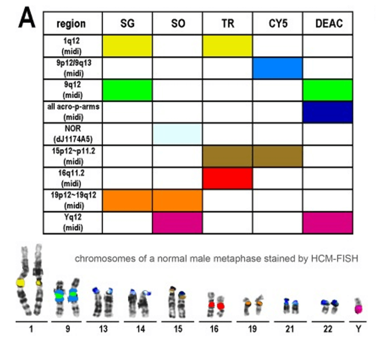 Fig 1. An example of a probe set for heterochromatin multicolor fluorescence in situ hybridization (HCM-FISH). (Bucksch M et al., 2012)
HCM-FISH Method
There is a close relationship between gene regulation, chromatin structure, and genome organization. Changing the position of a gene relative to the boundary between heterochromatin and euchromatin will affect its chromatin structure, which in turn affects its expression, manifested as transcriptional changes or phenotypic variations. The constitutive heterochromatin chromosomal region of mammals includes the central heterochromatin surrounding the centromeres, (sub)telomeres, and (peripheral) nucleolar organization (NOR). The HCM probe set is designed to target most acrocentric chromosomes, including chromosomes 13, 14, 15, 21, and 22. They contain several tandem copies of ribosomal RNA genes (specific to the nucleolar organization region (NOR)) on their short arms. The chromatin structural variation directed by the heterochromatin region is realized by the HCM-FISH probe set. These changes may include translocation, insertion, inversion, amplification, and formation of labeled chromosomes.
HCM-FISH Probe Set Accessible Area
The HCM probe set is designed to target most chromatin-concentrated regions on human chromosomes. In some literature, probe sets reported for HCM-FISH analysis cover chromosomes or regions, including the following items.
- Acrocentric short arms
- NORs
- Chromosomal 1/ 9/ 13/ 14/ 15/ 16/ 19/ 21/ 22/ Y
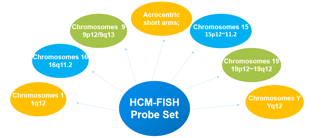 Fig 2. The classic accessible area of the HCM-FISH probe set.
Creative Bioarray provides a comprehensive probe customization service for the modification of probe sets in HCM-FISH analysis protocols. Our probe customization service can help customers expand analytical capabilities in experiments that visualize and analyze structural variation in heterochromatin regions on specific chromosomes. If you are interested in our services, please contact us for cooperation. We look forward to cooperating with you in the near future.
References
- Bucksch M, Ziegler M, Kosayakova N, et al. A new multicolor fluorescence in situ hybridization probe set directed against human heterochromatin: HCM-FISH[J]. Journal of Histochemistry & Cytochemistry, 2012, 60(7): 530-536.
- Singh P B, Belyakin S N, Laktionov P P. Biology and physics of heterochromatin-like domains/complexes[J]. Cells, 2020, 9(8): 1881.
All products and services on this website are only suitable for non-medical purposes.


