Gene Point Mutation
Creative Bioarray provides in-situ visual analysis services of gene mutations and SNPs based on specific target gene mutation site probes. Our service can use existing commercial specific mutation site probes or customized probes based on sites of interest. We provide one-stop analysis services from project design to output analysis, helping to test and analyze cancer-related gene mutation research related projects.
Gene Point Mutations Assay
The concept of precision medicine in oncology has emerged in the past decade and has solved the need for molecular characterization of individual tumors. This requires spatial analysis of tissue analysis in tumor samples to resolve inter-tumor and intra-tumor heterogeneity and to address the importance and regulation of subclonal mutations at the system level. In the past few years, many methods for spatially resolved transcriptomics have been developed. Among them, the targeted heavy multiple in situ methods includes unamplified single-molecule FISH (sm-FISH)-based methods and rolling circle amplification (RCA)-based methods. In situ-based methods have a subcellular resolution, and expression data can be directly correlated with morphology. The sm-FISH uses non-combined markers, composite markers, or their combination to achieve multiplexing. The main advantage is high sensitivity and can achieve a detection efficiency of close to 99%. The RCA-based method allows the use of 20x magnification to perform rapid imaging by detecting the intensity of the signal through amplification. Even in tumor tissue, the signal can be highlighted under the background of autofluorescence.
 Fig 1. In situ detection of KRAS mutations on prospective clinical samples with unknown mutation status. (Grundberg I, et al. 2013)
In situ Visualization Solution for Mutation Site Analysis
Probes based on a variety of specific methods have been commercialized. Our point mutation and SNP analysis in oncology is based on the mutation sites of genes with existing sequencing data, and the corresponding wild-type site probes are used for analysis. Take the in-situ analysis applied to the hybrid chain amplification reaction as an example. This service uses mutation-specific padlock probes, which have the same target sequence except that the last nucleotide at the 3' end varies by genotype, and performs in situ visual analysis of target gene mutations with wild-type padlock probes. Our service accepts testing items that currently have available commercial probes, and also accepts testing services based on custom probes. Customers can customize specific probes for mutation sites of genes supported by existing sequencing data according to their needs. The basic process of this service includes plan design (type of target gene of interest), sample pre-processing (quality control), probe selection (select commercial probes or custom), hybridization and imaging, and data analysis. After the customer submits information such as the target point of interest and sample type, we feedback the service plan and select commercial mutation site-specific probes or customized probes according to customer needs. After that, our experimental platform completes all analysis tests and submits data and analysis reports.
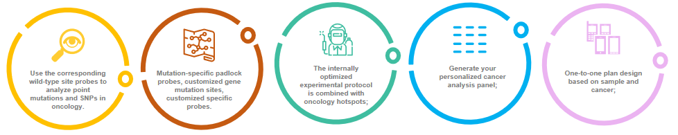 Fig 2. In-situ visualization service process for mutation site analysis.
A Case of Mutation Site Probe Set for Colon Cancer Analysis
- KRAS codons 12, 13 (G12S, G12R, G12C, G12D, G12A, G12V and G13D) and 61 (Q61H)
- EGFR (G719A, G719C, S768I and L858R)
- TP53 (S127F and P190S)
- Exons 11 and 15 of the BRAF gene
- Exons 1, 2, 9, 20 of the PIK3CA gene
- Other gene loci of interest
If you are interested in our service, please contact us for cooperation. We look forward to cooperating with you in the near future.
References
- Grundberg I, Kiflemariam S, Mignardi M, et al. In situ mutation detection and visualization of intratumor heterogeneity for cancer research and diagnostics[J]. Oncotarget, 2013, 4(12): 2407.
- Janiszewska M, Liu L, Almendro V, et al. In situ single-cell analysis identifies heterogeneity for PIK3CA mutation and HER2 amplification in HER2-positive breast cancer[J]. Nature genetics, 2015, 47(10): 1212-1219.
All products and services on this website are only suitable for non-medical purposes.


