Comet-FISH
Comet-FISH Technology
The genome integrity of organisms is constantly threatened by endogenous cellular metabolic processes and environmental factors. In order to quantify the level of DNA damage, a single-cell gel electrophoresis (comet) binding strand-specific FISH analysis method was established. Comet-FISH technology is a useful tool for detecting global and region-specific DNA damage and repair in a single cell. It is a combination of two mature methods, comet test (single cell gel electrophoresis) and FISH technology. Comet detection allows fragmented DNA to be separated from non-fragmented DNA, while FISH helps to detect specific labeled DNA sequences of interest, including entire chromosomes. The technical advantage lies in the quantification of low-level specific DNA damage in each strand of the selected sequence and the entire genome in a single cell with single-molecule sensitivity. By targeting different labels of specific FISH probes on both sides of a given sequence, DNA damage can be visualized as separate or adjacent or co-localized points under a fluorescence microscope. Therefore, the combination of these two technologies has been particularly applied to detect site-specific breaks in DNA regions related to the development of different diseases.
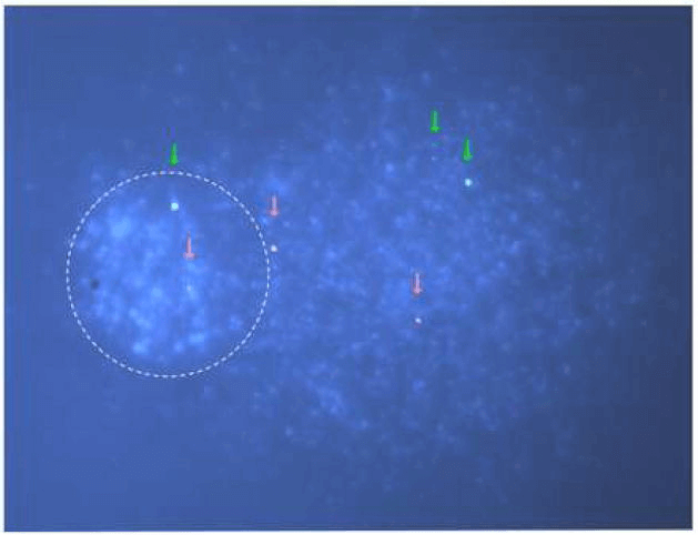 Fig 1. Representative image from a Comet-FISH assay. (Spivak G et al., 2009)
Comet Assay
Our Comet-FISH service can be used to compare and analyze the damage induction and repair of specific DNA sequences in genomes and single cells. The damage and repair status of the entire genome is quantified by the percentage of DNA in the comet tail. We provide customers with commercial or customized chain-specific FISH probes to analyze the damage of each chain of the specified sequence. Probes that target the 3'and 5'ends of the selected sequence are labeled with two different fluorophores. After comet FISH analysis, the two ends of the specified sequence are visualized as two different colored dots under a fluorescence microscope. The DNA damage event is indicated by the position of the probe, separated dots indicate damaged strands, and adjacent or co-located dots indicate complete strands.
With the development of this technology, various variants of probes have appeared, such as double-stranded fluorescent probes, fluorescent oligonucleotide probes, and padlock probes (rolling circle amplification). The advent of these probes aims to increase the resolution or scope of this technique. In addition to analyzing DNA damage that can be enzymatically or chemically converted into strand breaks, this technique has also been used to detect site-specific breaks in DNA regions associated with the development of different diseases.
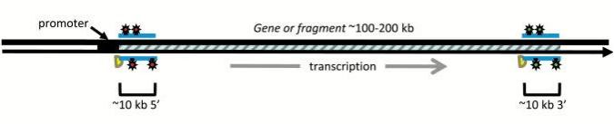 Fig 2. Probes for the transcribed and the non-transcribed strand are hybridized to separate groups of comet slides. (Spivak G et al., 2015)
Application
- Single cell DNA damage and repair assessment.
- Simultaneous analysis of GGR and TCR in cells exposed to low-dose genotoxic agents.
- Evaluation of special DNA damage events or DNA repair functions, such as transcription coupled repair (TCR) analysis.
- Analysis of DNA damage and repair in repair defect diseases.
- Assessment of DNA damage and repair caused by drug stimulation.
- The influence of site-specific genetic instability events on cell fate in cancer.
- Research the molecular mechanisms of various repair pathways and drug screening for the development of specific repair pathway inhibitors.
Creative Bioarray provides comprehensive FISH technology services and explores iterations of technology along the way. If you are interested in our services, please contact us for cooperation. We look forward to cooperating with you in the near future.
References
- Spivak G, Cox R A, Hanawalt P C. New applications of the Comet assay: Comet–FISH and transcription-coupled DNA repair[J]. Mutation Research/Reviews in Mutation Research, 2009, 681(1): 44-50.
- Schlörmann W, Glei M. Detection of DNA damage by comet fluorescence in situ hybridization[M]//DNA Repair Protocols. Humana Press, Totowa, NJ, 2012: 91-100.
- Spivak G. New developments in comet-FISH[J]. Mutagenesis, 2015, 30(1): 5-9.
All products and services on this website are only suitable for non-medical purposes.


