Creative Bioarray is an expert in analyzing chromatin interactions in the genome using 3D FISH technology. 2D FISH is used for mid-term research, while 3D FISH has been widely used to probe the relationship between the spatial organization of the genome and its interphase functions. 3D DNA FISH allows three-dimensional visualization of individual gene loci, subchromosomal domains and even entire chromosomes at all stages of the cell cycle. Our 3D DNA FISH service team is composed of scientists with rich experimental experience, providing you with convenient 3D DNA FISH solutions. We have established a mature program and can provide you with experimental reports and analysis results reports according to your requirements.
DNA FISH and Chromatin Interaction Analysis
The spatial organization of chromatin plays a very important role in the regulation of replication, repair and transcriptional activity. There are three main strategies for studying chromatin interactions in 3D genomes: methods based on high-throughput nuclear proximity connections, imaging tools, and computational/visualization methods. The first set of strategies covers methods based on chromosome conformation capture (3C). Imaging tools can directly visualize and quantify the spatial distance between genome sites, calculation methods are used to map global 3D genome structures at various scales, and visualization methods are used to visualize these data in a virtual 3D space with the help of different algorithms . Historically, co-association studies have been carried out by investigating dozens to hundreds of individual loci through FISH. Recently, powerful 3C-based high-throughput technologies have been developed, such as 4C (circularized chromosome conformation capture) and Hi-C, allowing the study of molecular crosstalk between thousands of different sites. 3D DNA FISH has become the main tool for analyzing the three-dimensional organization of cell nuclei, allowing three-dimensional visualization of individual gene loci, subchromosomal domains and even entire chromosomes at all stages of the cell cycle.
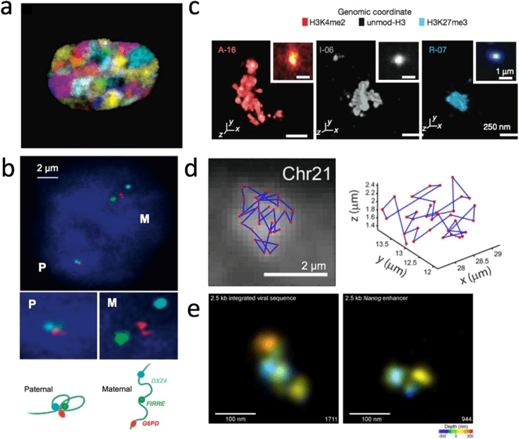 Fig 1. 3D genome visualization based on DNA-FISH methods. (Ma T, et al. 2018)
Services
One advantage of 3D DNA FISH is that it allows quantitative measurement of the three-dimensional distance between two target sites a and b in a single cell, allowing us to further estimate the distribution and degree of variation of the 3D distance between pairs of genomic sites. This method can provide valuable information about the basic configuration of chromatin fibers and can be used to infer possible models of chromosome folding. Our 3D DNA FISH service uses multicolor labeled probes complementary to specific target sites to hybridize to the genome and then uses a fluorescence microscope to image the sample. Finally, we use the image stack obtained by the confocal microscope to 3D model the FISH signal in the chromosome region and analyze the relative position of the hybridization probe in the reconstructed 3D image. In this way, 3D DNA FISH provides a powerful tool to ask how the tissue at a specific genomic locus changes in response to a stimulus.
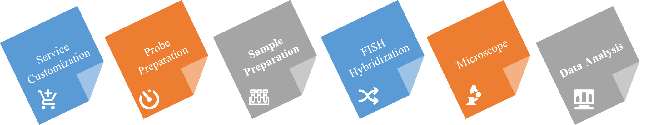 Fig 2. Our 3D DNA FISH service process.
Brief Steps
- DNA probe preparation and labeling procedures and nick translation.
It is produced by nick translation with amino-allyl adenosine diphosphate and chemical coupling with dye. This agreement is applicable to a range of cell types and various probes (BAC, plasmid, fosmids or whole chromosome paint);
- Cell fixation, pretreatment and permeabilization.
Determine the optimal sedimentation time and cell density according to the cell type, fix the cells of interest on a glass slide and perform permeabilization.
- 3D DNA FISH hybridization.
The sample DNA is denatured together with the directly labeled probe, and hybridized in a humidified chamber that is opaque.
- 3D DNA FISH detection.
- 3D DNA FISH microscope and analysis.
Creative Bioarray provides services for analyzing chromatin interactions in the genome using 3D FISH technology. Our DNA FISH experiment platform has the equipment and technology to carry out FISH analysis. You will benefit from our technical expertise and complete platform, and work with you to find the best solution to meet your needs.
If you are interested in our comprehensive 3D DNA FISH service, please contact us for cooperation. We look forward to cooperating with you in the near future.
References
- Bolland D J, King M R, Reik W, et al. Robust 3D DNA FISH using directly labeled probes[J]. Journal of visualized experiments: JoVE, 2013 (78).
- Jubb A, Boyle S. Visualizing Genome Reorganization Using 3D DNA FISH[J]. Methods in molecular biology (Clifton, NJ), 2020, 2148: 85-95.
- Ma T, Chen L, Shi M, et al. Developing novel methods to image and visualize 3D genomes[J]. Cell biology and toxicology, 2018, 34(5): 367-380.


