EGFRvIII in Glioblastoma
Creative Bioarray provides a visual analytics solution for EGFR in glioblastoma (GBM). This service is designed to help customers analyze the transcription level of the unique EGFR variable clips that exist in GBM. Rich FISH analysis service experience and professional team make us confident to provide you with industry-leading technical services.
EGFR and Glioblastoma
EGFR is a transmembrane receptor tyrosine kinase of the ERBB family. After binding to ligands (EGF, TGF-α, amphiregulin, β-cytokine, HB-EGF, epigen or epiregulin), EGFR forms homodimers or heterodimers with other ERBB family members. The dimerization of EGFR results in a phosphorylation event at its C-terminal tail, and the phosphorylated tail serves as a docking site for the signal protein containing the SRC homology 2 (SH2) domain for signal transduction. Many different EGFR mutations and rearrangements have been reported in cancer. But in different types of cancer, these events occur in different locations. Activating mutations in the kinase domain of EGFR are frequently detected in non-small cell lung cancer (NSCLC). These mutations include L858R in exon 21 and in-frame deletions in exon 19. A group of individual EGFR deletions and point mutations are often found in glioblastoma (GBM), including EGFRvI (N-terminal deletion), vII (deletion of exons 14-15), vIII (deletion of exons 2-7), vIV (deletion of exons 25-27), vV (deletion of exons 25-28). The detection of EGF rearrangement can help study its role in cancer.
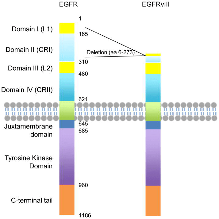 Fig 1. Functional domains of EGFR and EGFRvIII. (An Z, et al. 2018)
FISH Service of EGFRvIII in GBM
Among the EGFR mutants that GBM has discovered, EGFRvIII is the most common and is considered to represent a late event, which comes from the ancestral amplification of EGFR. Compared with EGFRWT, the lack of EGFRvIII results in a new connection site at the deleted fragment. This means that EGFRvIII has constitutive activity even though it does not contain a ligand binding domain. The role of EGFRvIII in the pathogenesis of GBM is still under study. We provide FISH-based visualization solutions for EGFRvIII in situ analysis. The probe design targets the flank of the deleted region of EGFRvIII. This set of probes covers the junction region of exons 1, 2, 7, 8, and 9, which can better realize the identification of mutations. This probe design can improve the specificity of EGFR analysis. This service can be carried out for FFPE tissue and tissue section samples. Our service process includes optional slice preparation, tissue permeabilization, FISH hybridization, imaging and data analysis. In the end, we will provide customers with at least 5 fields of raw data, analyzed data reports and experimental protocols.
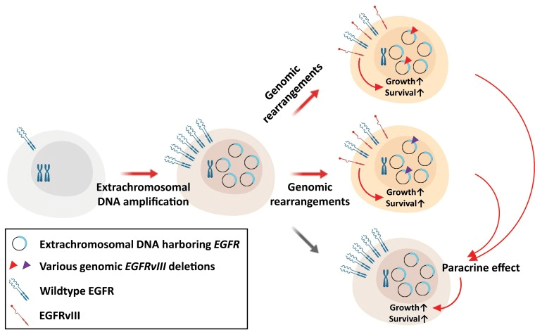 Fig 2. The possible mechanisms of tumor progression through genomic rearrangements of extrachromosomal DNA in glioblastoma. (Koga T, et al. 2019)
If you are interested in our FISH service , please contact us for cooperation. We look forward to cooperating with you in the near future.
References
- An Z, Aksoy O, Zheng T, et al. Epidermal growth factor receptor and EGFRvIII in glioblastoma: signaling pathways and targeted therapies[J]. Oncogene, 2018, 37(12): 1561-1575.
- Koga T, Chen C C, Furnari F B. When less is more: Gaining power through gene rearrangement of amplified EGFR[J]. Oncotarget, 2019, 10(22): 2116.
All products and services on this website are only suitable for non-medical purposes.


