PDX Models
Creative Bioarray has developed a visual analysis solution for the differentiation of gene expression of interest and species-specific expression in patient-derived xenograft (PDX) models. This service now realizes the visualization of target genes and the differentiation of species sources under the same morphological background. We choose species-specific commercial probe products or customized probes to achieve customer analysis goals. Our team of scientists can provide assistance in the design of the analysis plan. With the help of our experimental platform, you can visualize the personalized expression profile of PDX.
Gene Expression Analysis in PDX Models
Preclinical cancer models are the cornerstone of basic research and translational research. Existing models include cell culture models, animal models, and cell line-derived xenograft models. A PDX model refers to the implantation of biopsy specimens or surgically resected tumor samples into immunodeficient mice. It can also be produced by implanting malignant ascites-derived tumor cells or circulating tumor cells (CTC). At present, the PDX model is widely used as a preclinical cancer model and is considered to be superior to the cell culture model in terms of generalizing the histological characteristics, molecular characteristics, and intratumoral heterogeneity (ITH) of human tumors. Compared with cell culture in a petri dish, the PDX model better retains the biological and molecular characteristics and heterogeneity of patient tumors and is very useful for mechanism research, drug discovery, and other translational research in cancer research.
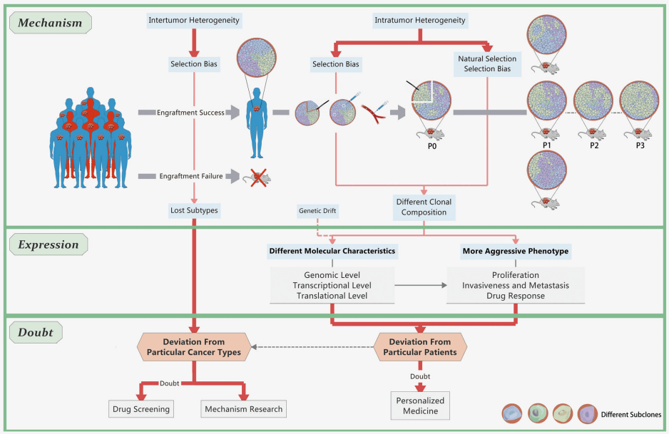 Fig 1. Infidelity of cancer cells in PDX models. (Shi J, et al. 2020)
Gene Expression and Location Analysis Service in the Humanized Model
Human tumor cells transplanted in PDX model mice grow in the mouse matrix environment, and these cells carry human tumors with the original genotype. However, the growth and metastasis of exogenous tumor cells supported by mouse tumor stroma require the differentiation of cell sources and types. These species-specific probes can be used to identify and quantify human and mouse-derived gene transcripts. The identification of the expression level in the study of the PDX model can be used to evaluate the spatial expression level in the tissue sample by in situ hybridization. At the same time, this part of the data can also be combined with the protein level to provide more evidence. Our gene expression and localization analysis are carried out by analyzing cells or slice samples derived from PDX models, using a wide range of commercial species-specific probes or customized species-specific probes. The analysis targets we can provide are known cancer biomarkers supported by sequenced data to distinguish the expression of donor and recipient sources.
Technical Features
Evaluate the expression of genes of interest in human tumor cells and donor stromal cells in the model, and distinguish expressions from different sources.
Perform specific gene target detection and visual analysis in a single sample with the same morphological background.
Simultaneously detect the spatial expression patterns of human and mouse specific targets.
Sample types include cells, tissue sections and tissue arrays.
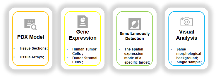 Fig 2. Our features and technical key points.
If you are interested in this in-situ visual analysis of gene expression in PDX models, please contact us for cooperation. We look forward to cooperating with you in the near future.
Reference
- Shi J, Li Y, Jia R, et al. The fidelity of cancer cells in PDX models: Characteristics, mechanism and clinical significance[J]. International journal of cancer, 2020, 146(8): 2078-2088.
All products and services on this website are only suitable for non-medical purposes.


