Chromosome-specific Probe
Microcloning Technology and Probe Customization
Chromosome microdissection (CMD) was first applied to the polytene chromosomes of Drosophila, and laser microdissection and microcloning techniques became effective and direct methods to isolate DNA from specific chromosomes and/or specific chromosomal fragments. The isolated DNA can be used for genome research, including the construction of genetic linkage maps and physical maps, preparation of chromosome mapping probes, and construction of chromosome-specific expression sequence tag libraries. For the preparation of a whole chromosome probe (WCP) or partial chromosome probe (PCP), common methods include microdissection or flow sorting. With the development and wide application of polymerase chain reaction (PCR), degenerate universal primers (DOP) are used to amplify microdissected or flow-sorted chromosomes, which makes microdissection applied in FISH.
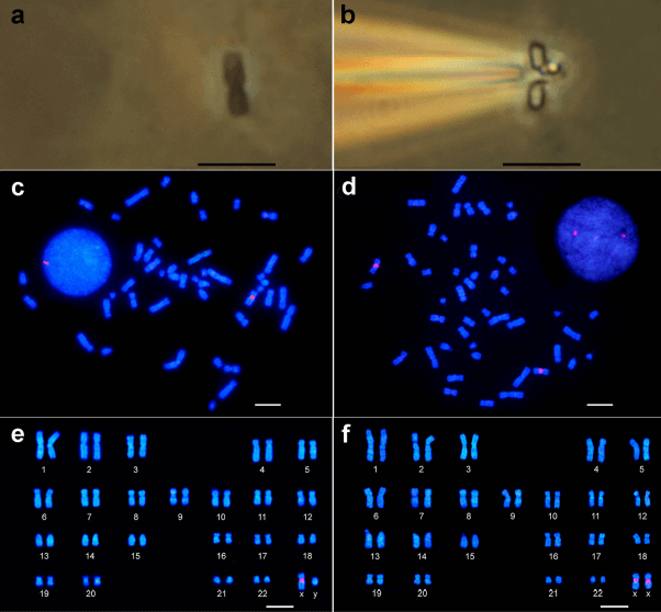 Fig 1. Microdissection procedure and FISH using chromosome-specific probe obtained via DOP-PCR. (Passamani P Z, et al. 2017)
Chromosome Microdissection and Probe Synthesis
Chromosome-specific probes such as WCP and PCP have been widely used in clinical diagnosis (prenatal and postnatal chromosome diagnosis), cancer genetics, and comparative cytogenetic research in various animal groups. Chromosome microdissection technology is currently mainly used in species with clearly identifiable target chromosomes or relatively simple chromosomes, such as polytene chromosomes and lamp-brush chromosomes. Our chromosome microdissection and probe preparation services are currently carried out mainly for samples of poultry origin, and we need to consult our experts for customized services for specific species.
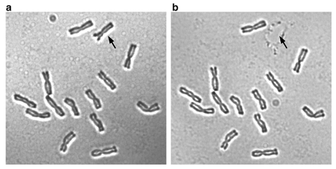 Fig 2. Microdissection of 1R chromosome of rye using a micromanipulator. (Zhang Y X, et al. 2016)
Our laboratory has the ability to prepare high-quality chromosome metaphases and conduct this service in an independent laboratory to avoid contamination and ensure product quality to the greatest extent. The basic process of this service is metaphase chromosome preparation, and then chromosome fragments are amplified in situ. Subsequently, the target area of the chromosome is microdissected using a microneedle coupled with a phase-contrast microscope. The obtained fragments are constructed into a DNA library and amplified by DOP-PCR, followed by probe preparation (choose appropriate dye labeling). The probes generated through this service can be used in metaphase FISH procedures to highlight these specific regions of metaphase chromosomes. Our probe customization service includes a series of internal quality control and hybridization verification steps to ensure the quality of the probe to the greatest extent.
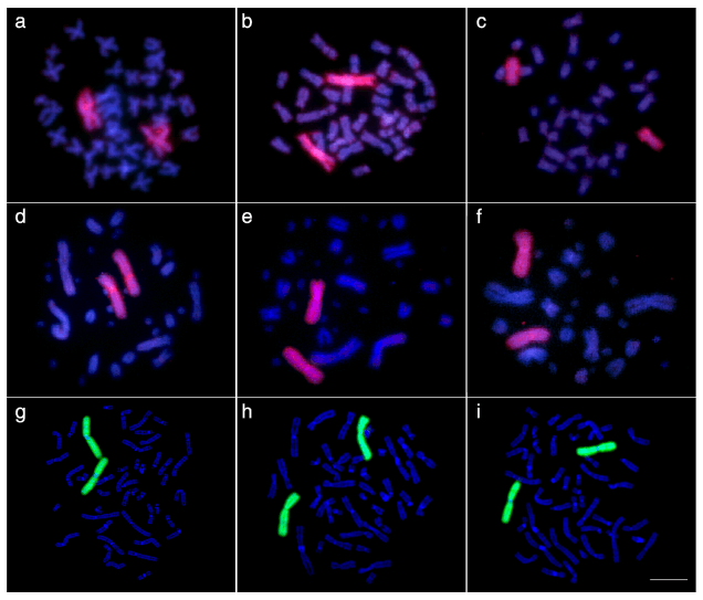 Fig 3. FISH with WCPs using probes prepared after microdissection procedures. (Al-Rikabi A B H, et al. 2019)
Application
- Used to efficiently construct chromosome-specific probes using relatively small fragments and low DNA copy numbers as templates;
- Used to generate probes for screening other chromosomal regions and analyzing structural and numerical abnormalities;
- Can be applied to many other organisms, not limited to human chromosomes;
Creative Bioarray provides customized services for microdissection of a single target chromosome and chromosome-specific probes. We have established an independent operation room for this service, and provide customers with high-quality customized probes through internally optimized quality control links and equipment. You will benefit from our expertise to meet your personalized analysis needs. If you are interested in our FISH service, please contact us for cooperation. We look forward to cooperating with you in the near future.
References
- Passamani P Z, Carvalho C R, Soares F A F. Protocol for chromosome-specific probe construction using PRINS, micromanipulation and DOP-PCR techniques[J]. Anais da Academia Brasileira de Ciências, 2017, 90: 41-47.
- Zhang Y X, Deng C L, Hu Z M. The chromosome microdissection and microcloning technique[M]//Plant Cytogenetics. Humana Press, New York, NY, 2016: 151-160.
- Al-Rikabi A B H, Cioffi M B, Liehr T. Chromosome Microdissection on Semi-Archived Material[J]. Cytometry Part A, 2019, 95(12): 1285-1288.
All products and services on this website are only suitable for non-medical purposes.


