Tissue Cryosections and FISH
Cryosections vs. Paraffin Sections in FISH
In situ hybridization (ISH) uses synthetic complementary RNA or DNA nucleotide probes to locate and detect sequences of interest in fixed cells, tissue sections, or entire tissue scaffolds. The preservation of target DNA or RNA in tissues requires the use of cross-linking fixatives, such as formalin. The use of formalin-fixed and paraffin-embedded tissue for DNA quantification will sacrifice tissue and relatively poor resolution to a certain extent. This process may hinder the probe's access to the target sequence. Some special steps are applied to the tissue section before the hybridization step in order to optimize the probe-based signal. Generally speaking, the processing of fixed tissue sections is a trade-off between increased hybridization signal and loss of tissue structure. The use of a strong cross-linking fixative (i.e. 10% formalin) and paraffin embedding can produce an excellent morphological background. However, due to the high temperatures used in the embedding process and the inability of the probes to approach the remaining RNA targets, there is significant RNA degradation and extensive cross-linking. In contrast, cryosections only provide a medium morphology, but the RNA remains intact, and the use of a weaker cross-linking fixative (i.e. 4% paraformaldehyde (PFA)) provides better probe access to the target RNA. In addition, cryosections of fresh tissues can shorten the events used in the sample fixation and embedding steps, shorten the cycle of analysis and detection, and provide the possibility of rapid turnover. Our platform provides comprehensive frozen section preparation and FISH analysis services for frozen sections.
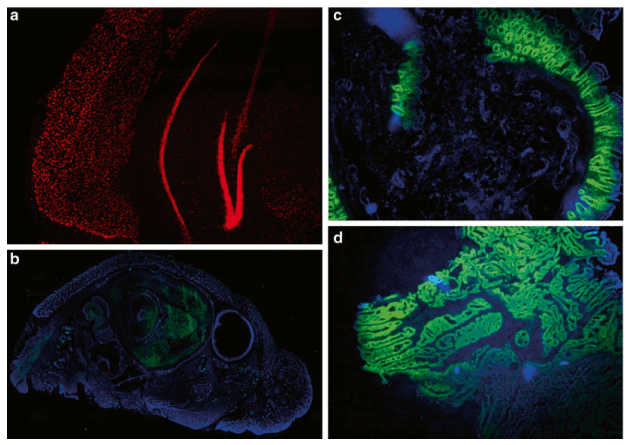 Fig 1. Different applications of microRNA FISH. (Silahtaroglu A et al., 2014)
DNA/RNA FISH in Tissue Cryosections
Our histology platform has established a complete frozen section preparation program, which can accept frozen section preparation services of fresh tissue samples and fixed tissue samples. We provide the preparation of frozen sections with a thickness of 8-12μm, which can be used for subsequent FISH hybridization analysis. For in situ visual analysis of cryosections, we provide solutions where the target is DNA or RNA. The difference between RNA itself and the characteristics of DNA (various types, instability, etc.), we provide solutions based on some special probes for analysis. For example, we provide the LNA-FISH protocol based on locked nucleic acid probes for the quantification and analysis of microRNAs in frozen sections. We provide LNA probe design and customization services, which can be used to carry out visual analysis on frozen sections. For more information, please consult our experts.
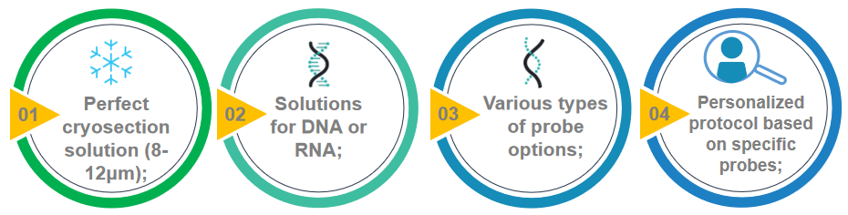 Fig 2. Key points of DNA/RNA FISH analysis in tissue cryosections.
Creative Bioarray provides DNA/RNA FISH analysis services on frozen sections. This service is for biological samples that are suitable for frozen sections, such as clinical samples. We provide a comprehensive one-stop service for frozen section preparation and FISH analysis, and you will benefit from our technical expertise. If you are interested in our FISH service, please contact us for cooperation. We look forward to cooperating with you in the near future.
References
- Silahtaroglu A. Fluorescence in situ hybridization for detection of small RNAs on frozen tissue sections[M]//In Situ Hybridization Protocols. Humana Press, New York, NY, 2014: 95-102.
- Asp J, Abramsson A, Betsholtz C. Nonradioactive in situ hybridization on frozen sections and whole mounts[J]. In Situ Hybridization Protocols, 2006: 89-102.
All products and services on this website are only suitable for non-medical purposes.


