Dual Color Break Apart Probe
Cytogenetic analysis shows that chromosomes that have been erroneously separated are hundreds of times more susceptible to the development of seven major types of structural aberrations. These chromosomal rearrangements include translocations, insertions, deletions, and the combination of chromosome fragmentation and classical non-homologous end-joining. Complex reorganization. For the lymphocyte progeny of the hematopoietic differentiation pathway, specific genome rearrangement/deletion may be a feature and necessary condition for many or even all differentiation lineages. In addition, to ending deletions, the observed rearrangement products require at least one DNA ligation event to occur between the ends of two broken chromosomes. Changes in chromosome structure are common in human cancers, and some genes are involved in many different translocations associated with cancer, so it is difficult to test every possible translocation using specific double fusion probes. A simple strategy is to use a dissociation probe to test whether the gene of interest is involved in the rearrangement. The break apart probe is designed to detect translocation, which is the most common genetic abnormality in cancer cells. With the continuous discovery of new gene rearrangements and the elucidation of their carcinogenic effects, translocation has become an important cancer biomarker. The occurrence of abnormal chromosome structure events brings about gene rearrangement, and molecular biology techniques such as FISH can determine the occurrence of these events.
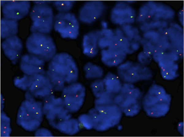 Fig 1. KMT2A (MLL) rearrangement analysis was performed using KMT2A dual color break apart probe. (Bao H, et al. 2019)
Break Apart Probe Customization Services
Our customized dual color break apart FISH probes are designed to help thousands of researchers quickly and reliably track gene translocations caused by abnormal chromosomal structures. The core of this service is to design sequence specific probes located on both sides of the gene for the fragments and genes of interest to customers and label them with two different fluorescent dyes. Our customized break apart probes can be used to detect translocation and rearrangement genes. When the translocation occurs, the two colors will separate. The dual color FISH probe produced by our gene break apart probe customization service allows the detection of two gene loci in a single sample, and each probe is assigned a different color dye. This is very useful for situations where multiple genes or regions need to be identified and can be used for the screening of genes of interest in cancer research.
Service Process:
- Submit target gene information;
- Choose the right combination of fluorescent dyes, or choose our preset combination;
- Sequence design and probe synthesis;
- Probe labeling and verification;
- Product delivery.
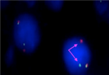 Fig 2. An example of a dual-color separation rearrangement probe used in gene rearrangement detection. (Thirunavukkarasu B, et al. 2021)
Creative Bioarray provides sequence-specific dual color break apart FISH probe customization to help customers analyze the gene rearrangement or chromosomal translocation region of interest. The core of this service is to design sequence-specific probes flanking the gene of interest for customers. You will benefit from our sequence design platform to provide you with a cost-effective FISH probe customized solution. If you are interested in our separation probe customization service, please contact us for customization. We look forward to cooperating with you in the near future.
References
- Bao H, Gao J, Chen Y H, et al. Rare myeloid sarcoma with KMT2A (MLL)-ELL fusion presenting as a vaginal wall mass[J]. Diagnostic pathology, 2019, 14(1): 1-6.
- Thirunavukkarasu B, Samanta J, Bhatia P, et al. De novo double-hit B-cell precursor leukemia/lymphoma-an unusual presentation as peritoneal lymphomatosis[J]. Autopsy and Case Reports, 2021, 11.
All products and services on this website are only suitable for non-medical purposes.


