CONTACT SUPPORT
Online Inquiry


Creative Bioarray's RNA ISH and IHC assay enables the visualization and quantification of CAR-NK presence and biomarker expression in processed tissue samples. We specialize in providing CAR-NK/Transgene/Cytokine/Biomarker quantification and statistical analysis services in a manner that is both high-quality and cost-effective. Our service can significantly expedite and validate the progression of your cell therapy developments.
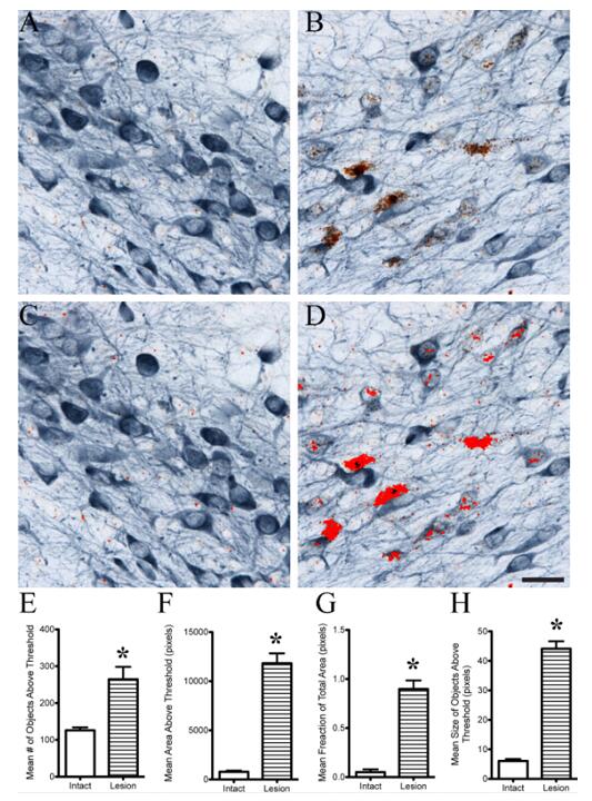 Fig 1. Semi-quantitative regional analysis of ISH signal in dual labeled (ISH-IHC) thick tissue sections.
A-D) Images were processed using ImageJ software to set the HSB index and hue was 0–128, brightness was 0–255 and saturation was 0–141. The pixels within the threshold limit are indicated by the bright red overlay. These setting allowed a distinction with fair accuracy between the Sprr1a ISH signal (brown) and the TH IHC signal (blue). The red areas in C and D are the pixels analyzed after the thresholds were set. E-H) After applying the threshold settings, image analysis was used to measure the average number of objects (E), mean area of pixels (F), mean fraction of the total area (G), and mean size of objects (H) (*p<0.05 compared to intact). With each measurement, the lesioned side is significantly greater than the unlesioned hemisphere. Scale bar: A-D = 40μm.
(1) Activated CAR-NK cell detection and quantification in intact fixed tissue
(2) Quantifiable assays to measure percentage of cells positive for CAR, transgene and biomarker expression
(3) Quantify CAR+ cell number and track persistence over time
Our customer service representatives are available 24hr a day! We thank you for considering Creative Bioarray as your CAR-NK/Transgene/Cytokine/Biomarker Quantification and Statistical Analysis partner.
References
SERVICES


Copyright © 2026 Creative Bioarray. All Rights Reserved.