FISH Analysis of Metaphase Chromosomes
Traditional FISH technology usually refers to the analysis of metaphase chromosomes, which is a different concept from interphase genetics. Traditional FISH is a single-cell technique, so it can identify low-frequency chromosomal abnormalities and determine which chromosomal abnormalities occur in the same or different clonal populations. Metaphase chromosomes are intact, so information about chromosomal homology abnormalities is retained. FISH analysis of metaphase chromosomes can help determine the organization of abnormal genomes after chromosome breaks and other types of complex genome rearrangements. The concept of metaphase FISH is differentiated from the later developed interphase FISH technology. This method can provide information about the abnormal chromosomal organization, balance rearrangement, or centromere, and other regions composed of highly repetitive DNA.
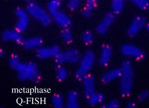 Fig 1. The PNA-labeled probe hybridizes with interphase cells. (Lai T P, et al. 2018)
Metaphase FISH Services
Metaphase FISH can also be used to determine the low-resolution distance between restriction markers. Compared with interphase, metaphase chromosomes have certain characteristics of intergenerational and genetic structure. Therefore, it can be used to analyze centromeres, telomeres, unique sequences, chromatids, and entire chromosomes. Our metaphase FISH analysis service provides multiple probe options to locate and analyze regions of interest on chromosomes at the same time. Customers can choose to analyze specific chromosomes or all chromosomes, mainly for the analysis of certain types of structures and characteristics of metaphase chromosomes, including analysis of centromere and telomere regions. The process includes experimental design, optional metaphase chromosome preparation, FISH probe customization, FISH hybridization, microscope services, and data services. For some FISH analyses related to mitosis, it can help determine the organization of abnormal genomes after chromosome breaks and other types of complex genome rearrangements.
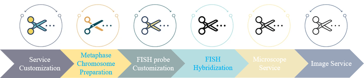 Fig 2. FISH analysis service of metaphase chromosomes.
Customers can mail simply processed cell or tissue samples to our laboratory. At the same time, our expert team will submit a personalized experimental plan according to your needs, and the specific details will be confirmed by you. After receiving the samples, we will complete simple sample processing, including optional metaphase chromosome preparation and sectioning. We provide probe synthesis and labeling services for FISH experiments, including optional nucleotide probes, peptide nucleotide probes (DNA and PNA), and select appropriate probe labeling schemes. After that, our FISH experiment platform will complete the hybridization experiment. After the high-quality data map is collected by the fluorescence microscope, it is handed over to the bioinformatics team for data collection and analysis. Finally, you will receive our complete experimental protocol, original data, and graphs for publication.
Our Features
- Our interphase and metaphase chromosome FISH service is a detailed service, you can choose a more affordable combination analysis service;
- Our experimental platform has a complete quality control protocol to ensure that customers' samples are maximized or get the best data;
- Our advanced laboratory services use industry-leading technology and equipment to ensure that customers receive the most optimized service;
Creative Bioarray provides a comprehensive FISH analysis service for metaphase chromosomes. You will benefit from our technology platform and special services. Our platform can complete all steps from sample pre-processing, and work with you to find a FISH solution that meets your needs. If you are interested in our metaphase chromosome FISH analysis service, please contact us for cooperation. We look forward to cooperating with you in the near future.
References
- Lai T P, Wright W E, Shay J W. Comparison of telomere length measurement methods[J]. Philosophical Transactions of the Royal Society B: Biological Sciences, 2018, 373(1741): 20160451.
- MacKinnon R N. Analysis of Chromothripsis by Combined FISH and Microarray Analysis[J]. Methods in molecular biology (Clifton, NJ), 2018, 1769: 53-77.
- Bustamante F O, Aliyeva-Schnorr L, Fuchs J, et al. Correlating the genetic and physical map of barley chromosome 3H revealed limitations of the FISH-based mapping of nearby single-copy probes caused by the dynamic structure of metaphase chromosomes[J]. Cytogenetic and genome research, 2017, 152(2): 90-96.


