FISH Service on Ultra-thin Cryosections
CryoFISH and cryoIF Technology
The visualization of cell structure and composition has become an important part of biological and medical science research. Immunofluorescence (IF) and FISH are powerful tools for visualizing the spatial relationship between proteins and nucleic acids in subcellular compartments. The resolution of the traditional laser scanning confocal microscope is up to ~200 nm. In order to use confocal microscopes to achieve higher spatial resolution, image contrast, and ultrastructure preservation, cryoFISH and cryoIF methods have been developed on ultrathin frozen sections of 100-200 nm. cryoFISH is a method of fluorescent in situ hybridization (FISH) on thin (~150 nm thick) frozen sections from fixed cells or tissues embedded in sucrose, which can be combined with immunoassays (IF) of other cellular components. Compared with whole-cell labeling methods, the main advantages of cryoFISH and cryoIF are improved spatial resolution, detection sensitivity (increased probe accessibility), and better image contrast through confocal microscopy. CryoFISH and cryoIF methods are usually used for samples that are fixed under the condition of preserving ultrastructure and are compatible with cell markers in the tissue environment.
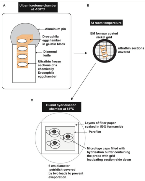 Fig 1. A schematic diagram of the key steps of an ultrathin section-based in situ hybridization method. (Rabouille C, et al. 2018)
Ultra-thin Frozen Sections and FISH Assay
The need for imaging of subcellular compartments and single molecules within cells has promoted the development of a wide range of sample preparation techniques and various microscope equipment to obtain images with higher spatial resolution. Our platform provides preparation services for ultra-thin sections based on sucrose embedding, and our platform provides ultra-thin sections with a thickness of about 150nm. Sections in this range can be used to distinguish cell submicroscopic structures and restore 3D cell structures. Our service can analyze the organelles containing nucleic acids, such as 28S ribosomes, mitochondria, etc. The basic process of the classic cryoFISH service includes sample pre-processing, ultra-thin section preparation, probe customization, FISH detection, imaging and data services. Our services can be carried out for cell and tissue samples. The samples need to be cross-linked in a fixative. Our experts will provide you with relevant technical guidance. In addition, the solutions of customers with cryoIF analysis needs need to be customized according to the target type.
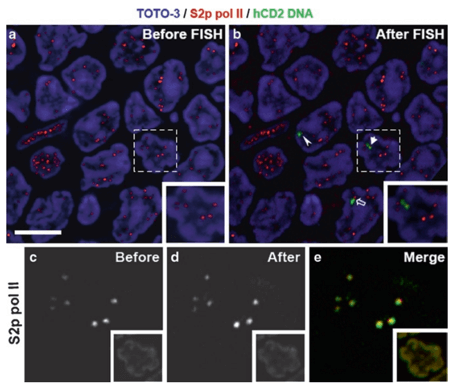 Fig 2. An example of locating molecules of interest in ultrathin sections. (Xie S Q, et al. 2010)
Technology Features
- The combination of confocal microscopy, electron microscopy, etc., and ultra-thin cryosections significantly improves the resolution, so that structures close to each other can be resolved as discrete bright spots under a high-contrast background, thereby improving the overall quantitative analysis.
- An additional advantage of the frozen section is its superior probe accessibility, especially when treated with mild detergent before IF/FISH.
Creative Bioarray provides cryoFISH analysis service based on ultrathin section preparation and optional cryoIF testing service. Our platform has established a laboratory with complete histological technology, which can provide high-quality FISH analysis and testing services. If you are interested in our cryoFISH service, please contact us for cooperation. We look forward to cooperating with you in the near future.
References
- Rabouille C. Detection of mRNA and Associated Molecules by ISH-IEM on Frozen Sections[M]//RNA Detection. Humana Press, New York, NY, 2018: 177-186.
- Xie S Q, Lavitas L M, Pombo A. CryoFISH: fluorescence in situ hybridization on ultrathin cryosections[M]//Fluorescence in situ Hybridization (FISH). Humana Press, Totowa, NJ, 2010: 219-230.
All products and services on this website are only suitable for non-medical purposes.


