Animal Preimplantation Embryos
Creative Bioarray offers 3D-FISH testing services, which can be used in research to analyze the correlation between nuclear organization and differentiation. Whole embryos can be analyzed by FISH with a specific fixation procedure that preserves the 3D shape of the embryo. Early embryonic development provides a particularly interesting but extremely complex system for studying how highly specialized gametes organize embryos, differentiate and generate new individuals after fertilization. Our services help customers analyze embryos and provide visualization analysis solutions for developmental analysis using embryos.
Visual Analysis on Mammalian Embryos
FISH for visualization of entire chromosomes or subchromosomal regions is a routine technique for chromosomal spread, as well as three-dimensional preservation of cells or tissues (3D FISH). FISH analysis performed on mammalian embryos can be used to study the nuclear organization of chromosomal and subchromosomal regions during the first developmental stage. FISH protocols for embryos preserved in three dimensions require a balance between adequate embryo permeability and maximal preservation of nuclear morphology. Embryos have greater depth compared to cells and are therefore not easily accessible for nuclear access with probes that visualize specific genomic regions by FISH. In early embryonic development of higher eukaryotes, the genome is organized in a highly spatially ordered manner, including individual genetic loci as well as complete chromosomes, organized into individual chromosomal regions (CTs). 3D-FISH techniques for 3D organs or 3D cultured tissues remain challenging, especially in the presence of large amounts of protein and RNA in embryonic cells at the preimplantation stage. Adaptation of the 3D-FISH protocol for embryonic specimens is challenging, and the harsh preprocessing brought about by the complex nature of the samples makes it necessary to balance hybridization efficiency with preservation of embryonic and nuclear morphology.
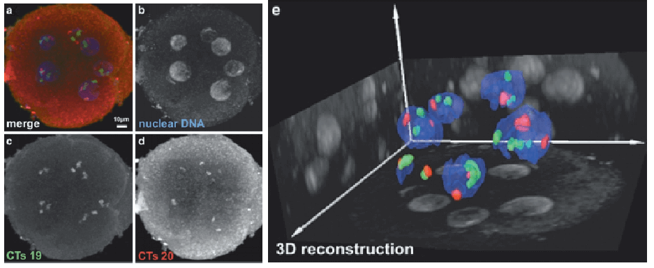 Fig 1. Bovine in vitro fertilized preimplantation embryo containing seven nuclei. (Daniela Koehler, et al. 2010)
Visualization Solution for Animal Embryos
After mammalian fertilization, the chromatin landscape inherited from both parental genomes and nuclear organization is reprogrammed. Tight regulation of nuclear organization is important for embryonic development and maturation. In cultured cells, their arrangement can be restricted according to their genomic content (gene density or duplication) or specific nuclear constraints (periphery or nucleolus). However, very little is known in the early stages of mouse embryonic development, especially about how and when the two parental genomes mix. Our FISH technical services provide 3D-FISH analysis solutions on archived embryos of mammals such as rats, mice, cattle, and pigs. We also provide analysis of transcripts for live cells of preimplantation embryos. The FISH protocol used by our platform has been optimized in-house and validated in embryos of the above species. We feature mapping probes using whole chromosomes as well as probes for subchromosomal regions, including commercial and custom probes.
 Fig 2. FISH detection of animal preimplantation embryos.
If you are interested in our service, please contact us for cooperation. We look forward to cooperating with you in the near future.
Reference
- Koehler, Daniela, et al. "FISH on 3D preserved bovine and murine preimplantation embryos." Fluorescence in situ Hybridization (FISH). Humana Press, Totowa, NJ, 2010. 437-445.
All products and services on this website are only suitable for non-medical purposes.


