Nucleus Separation in Paraffin-embedded Samples
The FISH technique, which uses in situ fluorescence to detect genetic abnormalities (such as translocations, amplification) in paraffin-embedded samples, is usually performed on tissue sections. However, the disadvantage of performing FISH on slices is that a large number of cells will lose part of the nuclear material (nuclear truncation) during the tissue slice process, resulting in an incomplete FISH labeling pattern (losing part of the target sequence used for hybridization). In addition, when it is necessary to distinguish one nucleus from another and evaluate the signal seen on different focal planes, FISH performed on slices is less effective (closely packed nuclei overlap each other). Therefore, the technology of FISH analysis of cell nuclei extracted from paraffin-embedded samples was born. The part of the surrounding cytoplasm preserved by the nucleus extracted from the paraffin-embedded tissue can be combined with FISH markers for immunocytochemical labeling of a series of cell markers (for example, lineage or proliferation). This allows the analysis of genetic abnormalities in specific cell populations. Cells isolated from paraffin sections can also be preserved in suspension to create special "tissue arrays" when needed for research using immuno-FISH technology (for example, to study new genetic abnormalities). We provide cell suspension preparation and FISH analysis services of paraffin-embedded samples such as biopsy tissues.
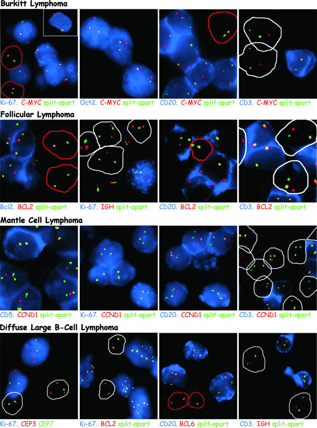 Fig 1. An example of immuno-FISH analysis of cells isolated from a paraffin-embedded lymphoma biopsy. (Mattsson G, et al. 2007)
Nucleus Suspension Preparation of Paraffin-embedded Samples
Reports for FISH analysis of suspensions prepared from paraffin-embedded samples usually refer to isolated cell nuclei, but such nucleus suspensions usually consist of intact or semi-intact cells. In this way, the cell nucleus can be used in traditional FISH analysis and ImmunoFISH procedures. The core of the technology consists of isolating nuclei from paraffin-sectioned or paraffin-embedded tissue samples and examining their status, as well as standard FISH analysis and ImmunoFISH analysis services. Nuclei isolation involves a series of steps of deparaffinization, precipitation, and separation, after which the cell suspension is made into special "sections". Such prepared suspensions can prepare "tissue arrays" for simultaneous analysis of multiple samples. Subsequent FISH analysis is adapted to standard FSIH protocols, with basic workflow including "slice" preparation, FISH hybridization, imaging, and data services. The ImmunoFISH service of our technical service platform combines immunohistological staining and FISH analysis to distinguish cells of different origins and types by identifying the properties of individual cells.
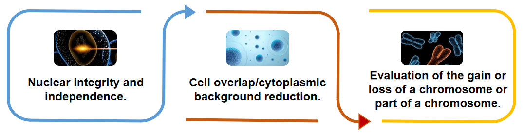 Fig 2. Features of FISH service carried out on isolated cell nuclei.
Technology Advantages
The main advantage of isolated nuclei obtained by preparing the suspension is that most of the nuclei are intact and can be clearly distinguished from their neighboring nuclei.
It is usually easier to evaluate FISH signal patterns in isolated cell nuclei than in tissue sections. This is because there are fewer nuclear truncation artifacts and the problems caused by excessive cell overlap/cytoplasmic background are less obvious.
Compared with the use of tissue sections, it is possible to more reliably evaluate and record the evaluation of complex abnormalities (for example, the gain or loss of chromosomes or parts of chromosomes) by the FISH program.
Creative Bioarray's FISH service can be used for a wide range of cytogenetic analyses. Our services can assist in the analysis of archived paraffin-embedded biopsy samples. If you are interested in our FISH service, please contact us for cooperation. We look forward to cooperating with you in the near future.
References
- Mattsson G, Tan S Y, Ferguson D J P, et al. Detection of genetic alterations by immunoFISH analysis of whole cells extracted from routine biopsy material[J]. The Journal of Molecular Diagnostics, 2007, 9(4): 479-489.
- Tan S Y, Mattsson G. ImmunoFISH on Isolated Nuclei from Paraffin-Embedded Biopsy Material[M]//Fluorescence in situ Hybridization (FISH). Humana Press, Totowa, NJ, 2010: 313-321.


