Chromogenic In Situ Hybridization
Chromogenic in situ hybridization (CISH) Technology
Chromogenic in situ hybridization (CISH) is a useful molecular biology analysis technique that can be used to assess gene amplification or deletion, chromosomal aneuploidy, or chromosomal translocation on tissue sections, and is often used in RNA in situ hybridization analysis. This technology uses a peroxidase-based color reaction to determine the amplification of the gene of interest (GOI) and is a relatively new technical method. Multiplex FISH of genes allows simultaneous detection of multiple targets using dual-color probes.
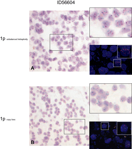 Fig 1. Chromogenic in situ hybridization (CISH) and repeated fluorescence in situ hybridization (FISH) analysis in tumor ID56604 (OIII). (Lass U et al., 2013)
CISH Assay
This powerful technique uses a novel probe design strategy and hybridization-based signal amplification system to simultaneously amplify the signal and suppress the background, enabling efficient single-molecule visualization in single cells. A large number of commercial probes exist for CISH analysis that can be applied to cells and fixed tissues to provide knowledge of the correlation between specific gene expression and tissue compartments and cell morphology.
CISH v.s.FISHs
Compared with FISH, it has been reported that the concordance rate of results between CISH and FISH is about 90%, indicating that CISH is a technique comparable to FISH. Although CISH is sometimes less sensitive for quantification of low-level expression. CISH has some features over FISH in the reagents and equipment it uses. For example, CISH is cheaper and easier to use because it uses brightfield microscopy instead of fluorescence microscopy; CISH reagents are more stable than FISH reagents, so the same sample can be examined multiple times. Additionally, FISH requires a high-resolution digital camera to capture photomicrographs of the sample before the fluorescence has subsided.
Another advantage of using brightfield microscopy for CISH is that whole tissue or cell samples can be visualized and cell morphology assessed by CISH. There are also differences between the two techniques in the probes used and in the overall approach. FISH can be performed using direct labeling (a fluorescent dye is attached to the probe) or indirect labeling (the probe is labeled with biotin or digoxigenin and then detected with fluorescently labeled streptavidin or antibody, respectively). In contrast, CISH is performed using indirect labeling.
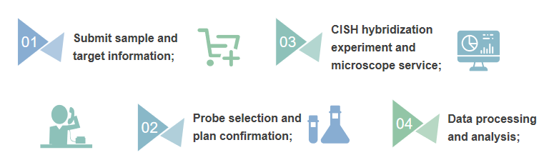 Fig 2. Flow chart of CISH.
- Sample quality control report
- Probe information report
- Experimental protocol and raw data report
- Data analysis reports and graphs that can be used for publication
Creative Bioarray offers probe products that can be used for CISH analysis. Our products go through rigorous quality control and validation steps, allowing you to harvest high-quality maps. We look forward to your choice of our FISH analysis services as an alternative to CISH analysis. Please contact us for consulting services and look forward to cooperating with you in the near future.
References
- Lass U, Hartmann C, Capper D, et al. Chromogenic in situ hybridization is a reliable alternative to fluorescence in situ hybridization for diagnostic testing of 1p and 19q loss in paraffin‐embedded gliomas[J]. Brain Pathology, 2013, 23(3): 311-318.
- Di Palma S, Collins N, Faulkes C, et al. Chromogenic in situ hybridisation (CISH) should be an accepted method in the routine diagnostic evaluation of HER2 status in breast cancer[J]. Journal of clinical pathology, 2007, 60(9): 1067-1068.
All products and services on this website are only suitable for non-medical purposes.


