Creative Bioarray provides usable in-situ visualization analysis solutions for 3D cell culture models. FISH allows the visualization of specific regions of the genome for spatial mapping. FISH in 3D culture is technically more challenging and practical than single culture. We provide analysis solutions for mammary gland acinar models and multicellular tumor spheres (MCTS) models.
Solid Tumors and 3D Cell Culture
The importance of the spatial positioning of DNA for the correct function of the genome has been paid more and more attention. It has been observed that the spatial positioning of the genome will change during diseases such as cancer. Most gene mapping studies are performed on cultured cells, but the organization of the genome may be different in the tissues. The three-dimensional (3D) culture model system provides a powerful tool for studying the contribution of tissues to gene expression and tissues. Many cell types have been grown under various 3D culture conditions, and various types of in vitro 3-D culture systems have been developed to summarize cancer growth conditions in vivo. The cancer 3-D culture method is designed to better retain the biological characteristics of the original tumor than the traditional 2-D monolayer culture, including tumor-derived organoids, tumor-derived spheroids, organotypic multicellular spheroids, and multicellular tumor spheroids. For example, the more widely used mammary gland acinar models not only resemble the breast morphology in vivo but also reproduce the function of the glands, which can be used for the research of various problems in the field of cancer.
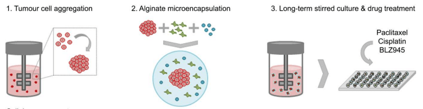 Fig 1. The basic process of tumor 3D model construction. (Rebelo S P, et al. 2018)
FISH Services on 3D Cell Model
Our FISH service can visualize specific regions of the genome in cell models and map genomic loci in interphase cells. The basic process of the service is sample pretreatment (fixation, permeabilization, and DNA denaturation), probe selection and customization, FISH hybridization, imaging and data analysis. Compared with 2D cultured cell models, 3D cultures are thicker than standard single cultures and contain an extracellular matrix, which increases the difficulty of probe penetration, and 3D cultures are more fragile and more difficult to handle and manipulate. Our internally optimized experimental protocol has improved the problem through multiple steps, including optimizing permeabilization conditions, increasing denaturation time, and increasing temperature. Our experimental platform has carried out this test service on the 3D cultured breast acinar model. In addition, the multicellular tumor spheres in the tumor 3D culture cell model are established from cancer cell lines in a traditional medium supplemented with FBS, similar to traditional 2-D culture. Methodologically, it can be regarded as an extension of the standard two-dimensional culture of cancer cell lines. Compared with other 3-D systems, the advantages of cell clonality, easy maintenance and simplicity of genetic manipulation make this method a suitable tool for high-throughput drug testing. We also provide testing services for this model. For more information, please consult our experimental team, we will customize the plan for your research one-to-one.
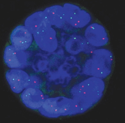 Fig 2. FISH on 3D culture structures. (Meaburn K J, et al. 2010)
Service Features
Optimized experimental protocol. We improve the analysis effect by increasing the accessibility of probes.
Our testing services can be carried out for 3D cultured breast acinar models and MCTS models.
We provide one-to-one customized solutions to provide customers with the best solution.
Creative Bioarray provides visual analytics solutions for some 3D cultured cell models. Through the optimization of the classic FISH solution, the visual analysis problem in the 3D model is solved, and the solution is customized for customers. If you are interested in our FISH service, please contact us for cooperation. We look forward to cooperating with you in the near future.
References
- Rebelo S P, Pinto C, Martins T R, et al. 3D-3-culture: A tool to unveil macrophage plasticity in the tumour microenvironment[J]. Biomaterials, 2018, 163: 185-197.
- Meaburn K J. Fluorescence in situ hybridization on 3D cultures of tumor cells[M]//Fluorescence in situ Hybridization (FISH). Humana Press, Totowa, NJ, 2010: 323-336.


