CONTACT SUPPORT
Online Inquiry


The rapid advancements in the field of cell and gene therapy have revolutionized the treatment of various genetic and degenerative diseases. As researchers and scientists strive to develop innovative methods to improve the efficacy and safety of these therapies, fluorescence in situ hybridization (FISH) has emerged as a powerful tool for the visualization and analysis of genetic material within cells and tissues.
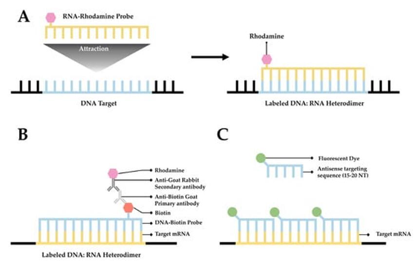 Fig. 1 A simplified diagram depicting FISH. (Tingey M, et al., 2022)
Cell and gene therapy have garnered significant attention due to their potential to treat a wide range of diseases, including cancer, genetic disorders, and autoimmune conditions. Chimeric Antigen Receptor (CAR) T-cell therapy has shown promise in the treatment of certain types of leukemia and lymphoma. AAV (adeno-associated virus)-based therapies are effective for some types of genetic diseases. Currently, cell and gene therapy research is focused on optimizing the delivery, targeting, and efficacy of gene therapy. As this field continues to evolve, there is a growing need for advanced tools and methods to assess the biodistribution of therapeutic genes and cells within organisms.
Biodistribution (BD) studies play a pivotal role in evaluating the dissemination and localization of gene therapy vectors and cellular therapeutics within the body, according to guidelines for biodistribution studies developed by different regulatory agencies and international scientific organizations. In a guidance document from the Food and Drug Administration (FDA), the BD study is viewed as one of the considerations for the determination of the cell fate, which refers to the conditions of cells after administration determined by assessment of survival/engraftment and distribution, differentiation/integration, and tumorigenicity of cells. Accurate assessment of biodistribution is crucial for ensuring the effective targeting of therapeutic genes and cells to the intended tissues and organs. Furthermore, it aids in monitoring potential off-target effects and the overall safety profile of the treatment.
FISH is a molecular cytogenetic technique that enables the visualization and mapping of specific DNA or RNA sequences within cells and tissues. The technique utilizes labeled nucleic acid probes that hybridize to complementary target sequences, allowing for the precise localization and detection of genetic material. The history of FISH dates back to the 1980s. Since then, it has undergone remarkable advancements, including the development of RNAscope, Basescope, and miRNAscope, which can accurately detect very short sequences.
Applications of FISH in gene and cellular localization
FISH technology plays a crucial role in the precise localization of therapeutic genes, vectors, and cellular components within target tissues. By leveraging FISH probes tailored to specific genetic sequences, researchers can visualize the spatial distribution of genetic material, providing invaluable insights into the intracellular and intravascular localization of gene therapy vectors and transduced cells. This capability is instrumental in optimizing the delivery and targeting of gene therapy products, ultimately enhancing their therapeutic efficacy.
Applications of FISH in gene diagnosis and gene efficacy assessment
FISH facilitates the visualization of therapeutic gene vectors, such as AAV and lentivirus, within tissues by employing specific probes designed to target and identify the transferred genetic material. This enables researchers to trace the spatial distribution of the therapeutic genes and precisely determine their localization within the target tissues.
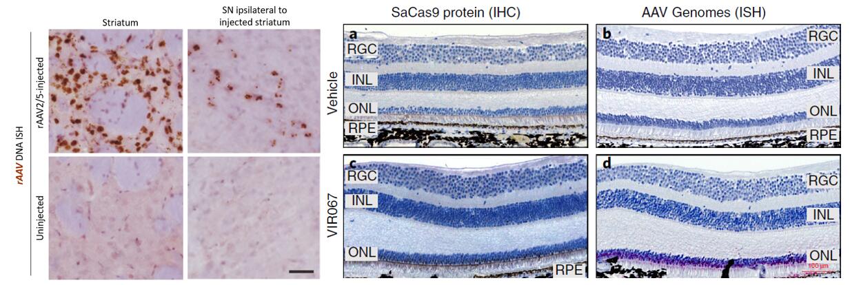 Fig. 2 Detection of rAAV viral DNA injected in the rat striatum using the RNAscope technique (Grabinski TM, et al., 2015); The distribution of SaCas9 in the retina was examined using RNAscope (Maeder ML, et al., 2019).
FISH can be used to quantitatively assess the expression of therapeutic genes within the target tissues, providing valuable insights into the extent and efficiency of gene delivery. By detecting and counting the copies of the inserted gene sequences, FISH enables the measurement of transgene expression levels, thereby contributing to the evaluation of gene therapy efficacy.
| Targets | Detection Methods | Purposes |
|---|---|---|
| AAV (adeno-associated virus) gene vectors | RNAscope, BaseScope | Visualizing and quantitative analysis of AAV vectors and transgene expression within target cells. |
| Lentivirus gene vectors | RNAscope, BaseScope | Detection and localization of vectors and incorporated transgenes within host cells. |
| siRNA (small interfering RNA) | miRNAscope | Visualization and quantitative analysis of siRNA uptake and intracellular localization. |
| miRNA (micro-RNA) | miRNAscope | Visualization and quantitative analysis of miRNA localization and expression within cells. |
| ASO (antisense oligonucleotide) | miRNAscope | Visualization and quantitative analysis of the intracellular localization and targeting of ASO molecules. |
Application of FISH in cell therapy development
FISH allows for the visualization and precise localization of transplanted cells within living organisms. By utilizing cell-specific FISH probes, researchers can track the cellular biodistribution and monitor the spatial distribution of transplanted cells within the target tissues over time.
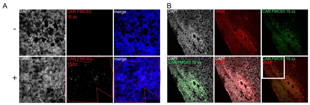 Fig. 3 In situ detection of anti-CD19-CAR T cells in mouse xenografts of NSG mice after anti-CD19 CAR T cell infusion (Eichholz K, et al., 2021).
FISH technology enables the assessment of cell engraftment, retention, and proliferation within the target tissues. Researchers can analyze the dynamics of transplanted cell localization and integration, providing critical insights into the efficacy of cell therapy interventions.
| Targets | Detecting Methods | Purposes |
|---|---|---|
| CAR-T (Chimeric Antigen Receptor T-Cell) | RNAscope, BaseScope | Tracking the localization and quantitative analysis of CAR-T cells within the tumor microenvironment. |
| CAR-NK (Chimeric Antigen Receptor Natural Killer Cell) | RNAscope | Visualization and quantitative analysis of CAR-NK cell localization and dynamics. |
| CAR-Macrophage (Chimeric Antigen Receptor Macrophage) | RNAscope | Assessing the localization and quantitative analysis of CAR-Macrophage cells within the immune microenvironment. |
References
SERVICES


Copyright © 2026 Creative Bioarray. All Rights Reserved.